Spinal Stenosis Symptoms, Diagnosis & Treatment Miami Neuroscience Center
The central canal is the portion of the ventricular system of the central nervous system that is specific to the spinal cord. It originates when the embryonic neural tube folds together forming a lumen. It carries cerebrospinal fluid in early years but is often partially or wholly obliterated in adults. The central canal is generally oval in.
Spinal cord Encyclopedia Anatomy.app Learn anatomy 3D models, articles, and quizzes
Hydromyelia refers to an abnormal widening of the central canal of the spinal cord. This widened area creates a cavity in which cerebrospinal fluid (commonly known as spinal fluid) can build up. As spinal fluid builds up, it may put abnormal pressure on the spinal cord, and damage nerve cells and their connections.

The structure of the spinal cord Spinal cord, Nervous, Muscle anatomy
Human Anatomy (OERI) 12: Central and Peripheral Nervous System
Central Canal Stenosis Definition Spine Info
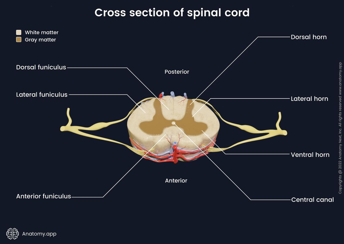
The central canal, also known as the central spinal canal or the vertebral canal, is a cylindrical channel that runs the length of the spinal cord. It is located at the center of the vertebral column and is surrounded by the vertebral arches, which are bony structures that form the spinal column's protective framework.
Spinal Cord
The spinal cord is part of the central nervous system (CNS). It is situated inside the vertebral canal of the vertebral column. During development, there's a disproportion between spinal cord growth and vertebral column growth. The spinal cord finishes growing at the age of 4, while the vertebral column finishes growing at age 14-18.

Histology of the Spinal Cord YouTube
Central Cord Syndrome. Central cord syndrome (CCS) is an incomplete traumatic injury to the cervical spinal cord - the portion of the spinal cord that runs through the bones of the neck. This injury results in weakness in the arms more so than the legs. The injury is considered "incomplete" because patients are usually not completely.

front
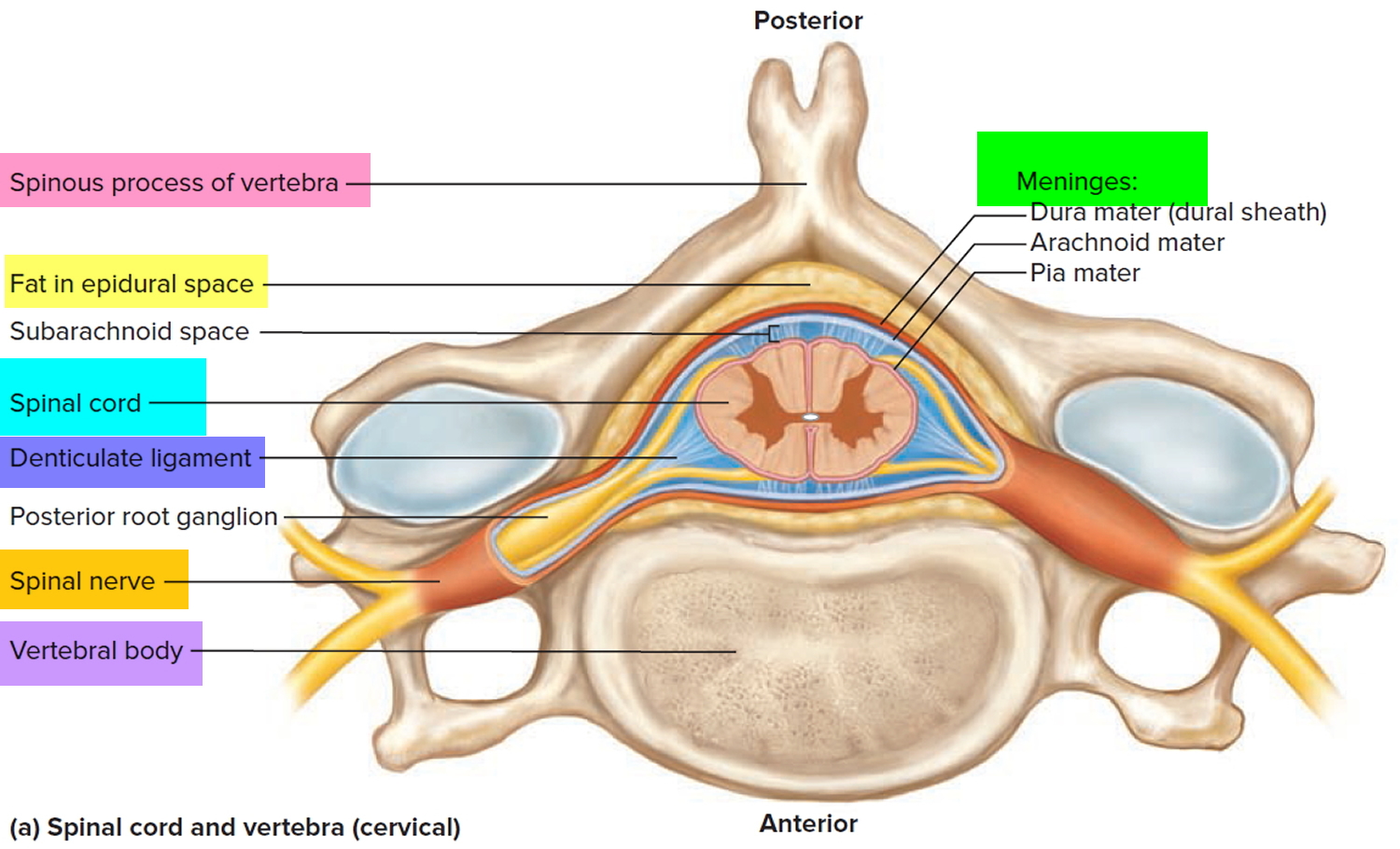
The spinal cord is part of the central nervous system (CNS), which extends caudally and is protected by the bony structures of the vertebral column. It is covered by the three membranes of the CNS, i.e., the dura mater, arachnoid and the innermost pia mater. In most adult mammals it occupies only the upper two-thirds of the vertebral canal as the growth of the bones composing the vertebral.
PPT Exercise 19 PowerPoint Presentation, free download ID6084979
Anatomy Answer: The central canal is a fluid-filled space in the spinal cord that has a protective function and allows for nutrient transport. The ventricles in the brain are filled with a high salinity solution called cerebrospinal fluid. This CSF is produced by ependymal cells in the choroid plexus, which lines the ventricles in the brain.
PPT The spinal cord provides a vital link between the brain and the rest of the body, and yet
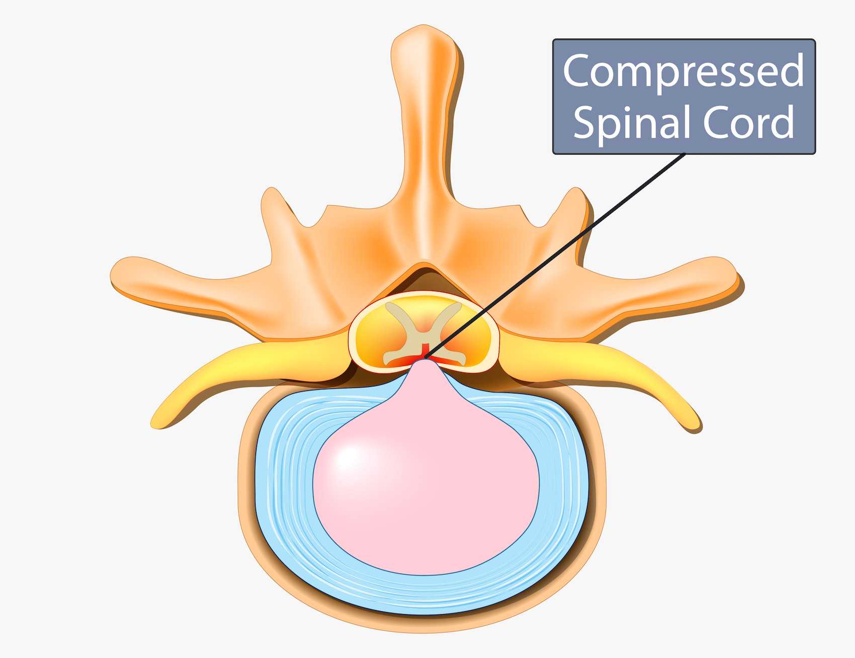
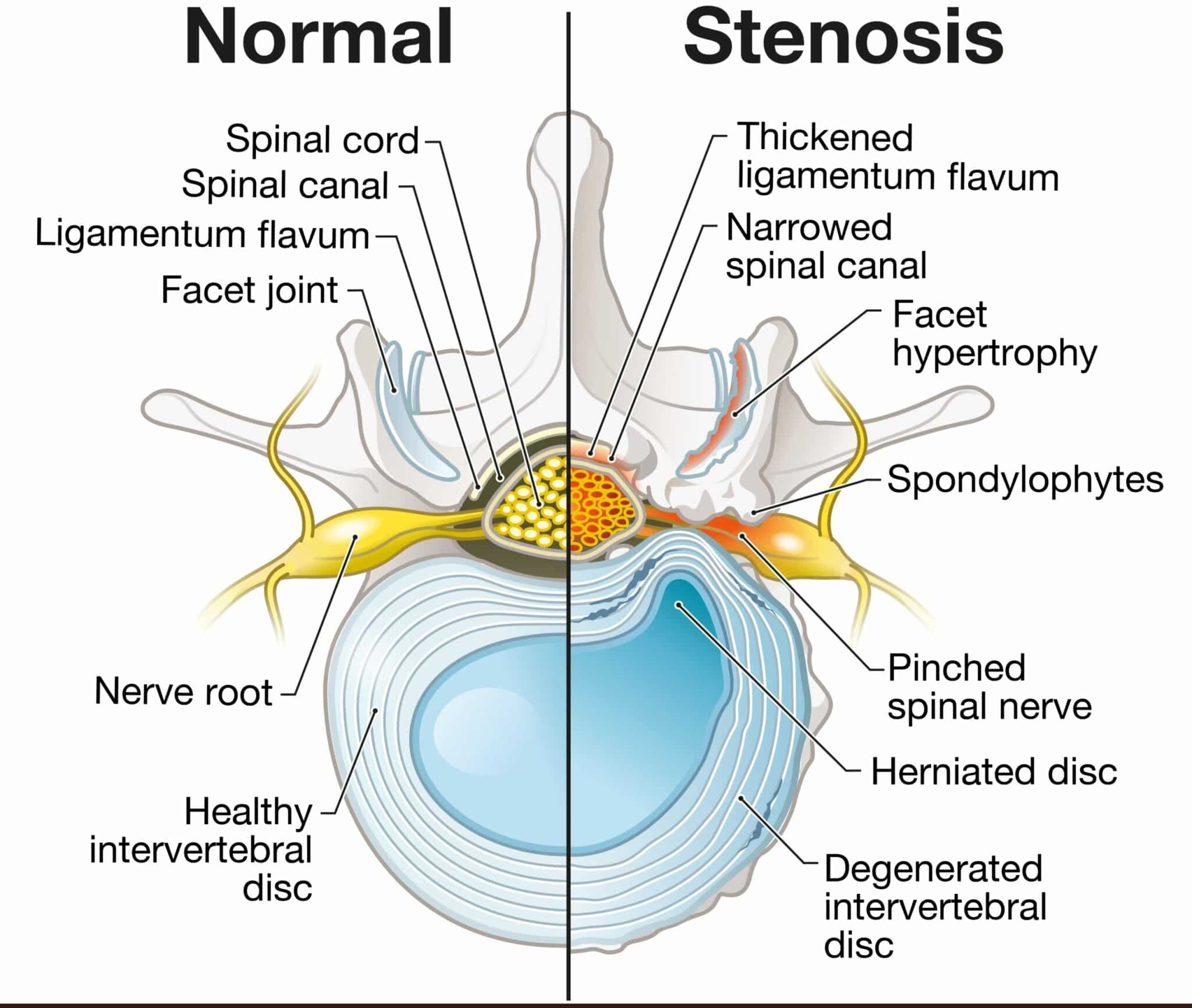
Overview Spinal stenosis happens when the space inside the backbone is too small. This can put pressure on the spinal cord and nerves that travel through the spine. Spinal stenosis occurs most often in the lower back and the neck. Some people with spinal stenosis have no symptoms. Others may experience pain, tingling, numbness and muscle weakness.
Central canal definition — Neuroscientifically Challenged
Derived from the primitive neural tube, the central canal encompasses an internal system of cerebrospinal fluid (CSF) cavities that include the cerebral ventricles, aqueduct of Sylvius, and fourth ventricle [ 1 ].
PPT Spinal Cord PowerPoint Presentation, free download ID6573953
Prominent central canal: This refers to a slightly expanded central canal filled with cerebrospinal fluid (CSF) without any spinal cord signal abnormality or enhancement. Cystic spinal cord neoplasm: The imaging hallmarks are cord signal abnormality, mass effect, contrast enhancement, and associated with neurological symptoms. 1 Syringohydromyelia:
Spinal Cord Anatomy Parts and Spinal Cord Functions
Cerebrospinal fluid (CSF) is a clear, colorless plasma-like fluid that bathes the central nervous system (CNS). Cerebrospinal fluid circulates through a system of cavities found within the brain and spinal cord; ventricles, subarachnoid space of the brain and spinal cord and the central canal of the spinal cord.

Central Canal Stenosis Symptoms, Causes and Treatment Spinal Backrack
The central canal (also known as spinal foramen or ependymal canal [1]) is the cerebrospinal fluid -filled space that runs through the spinal cord. [2] The central canal lies below and is connected to the ventricular system of the brain, from which it receives cerebrospinal fluid, and shares the same ependymal lining.

Spinal Cord Sectional Anatomy Spinal Cord Anatomy, Spinal Cord Injury, Gross Anatomy, Human Body
The Human Central Canal of the Spinal Cord: A Comprehensive Review of its Anatomy, Embryology, Molecular Development, Variants, and Pathology . Authors Erfanul Saker 1 , Brandon M Henry 2 , Krzysztof A Tomaszewski 2 , Marios Loukas 1 , Joe Iwanaga 3 , Rod J Oskouian 4 , R Shane Tubbs 5 Affiliations
Internal Anatomy Of Spinal Cord Anatomy Book
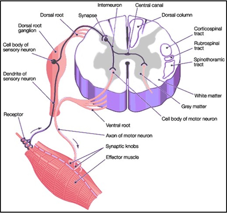
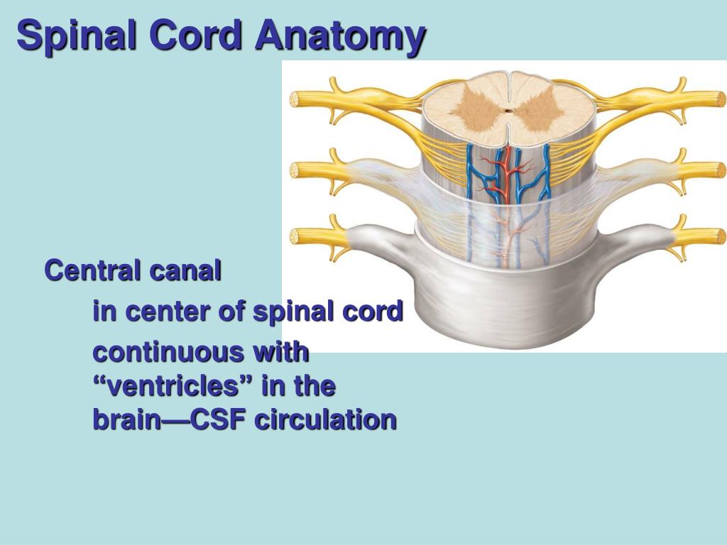
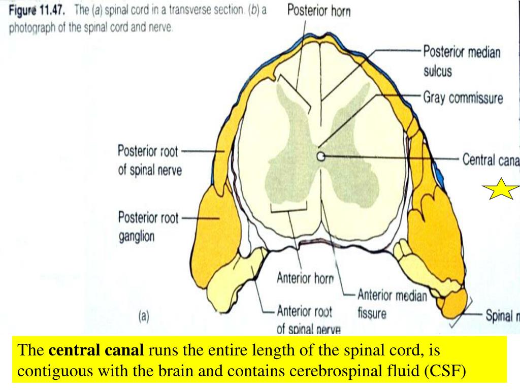
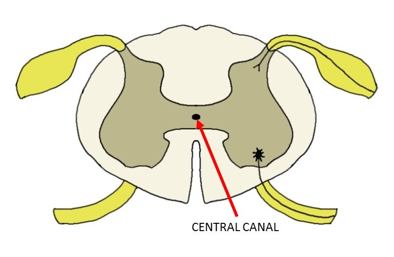
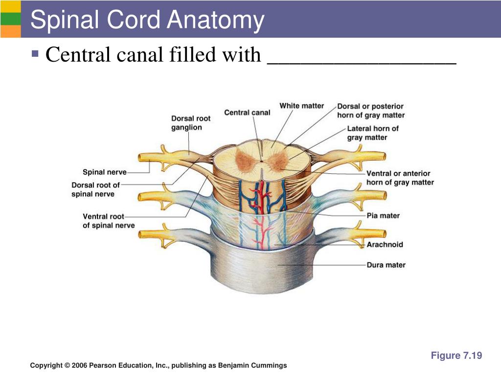
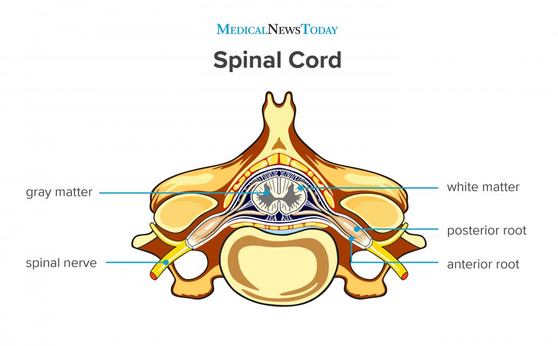
In the central region of the spinal cord, a central canal is the continuation of the fourth ventricle of the brain and contains cerebrospinal fluid (CSF). Surrounding the central canal, a horizontal line of gray matter called the gray commissure connects the left and right sides of the spinal cord.

Anatomy of the spinal cord. Download Scientific Diagram
The vertebral canal, otherwise known as the vertebral cavity or spinal cavity, is an anatomical space formed by the vertebral column that stores an integral portion of the central nervous system: the spinal cord and the spinal nerve roots branching off the spinal cord bilaterally.
