
MTA in Front of MRI Machine in Radiology Stock Image Image of people, machine 33010943
The Radiology Assistant : Epilepsy - Role of MRI Epilepsy - Role of MRI Laurens De Cocker, Felice D'Arco and Philippe Demaerel and Robin Smithuis Publicationdate 2012-09-01 In many patients with epilepsy antiepileptic drug treatment is unable to control the seizures.

Radiography Exam New England School of Dental Assisting
Objective The aim of the study is to visually rate major forms of dementia using global cortical atrophy (GCA), medial temporal lobe atrophy (MTA), and Fazeka's scales and Koedam's score using magnetic resonance imaging (MRI). The purpose is to correlate the visual rating scales (VRS) with severity of dementia. Materials and Methods Thirty patients fulfilling DSM 5 (Diagnostic and.
Registered Radiologist Assistant ARRT
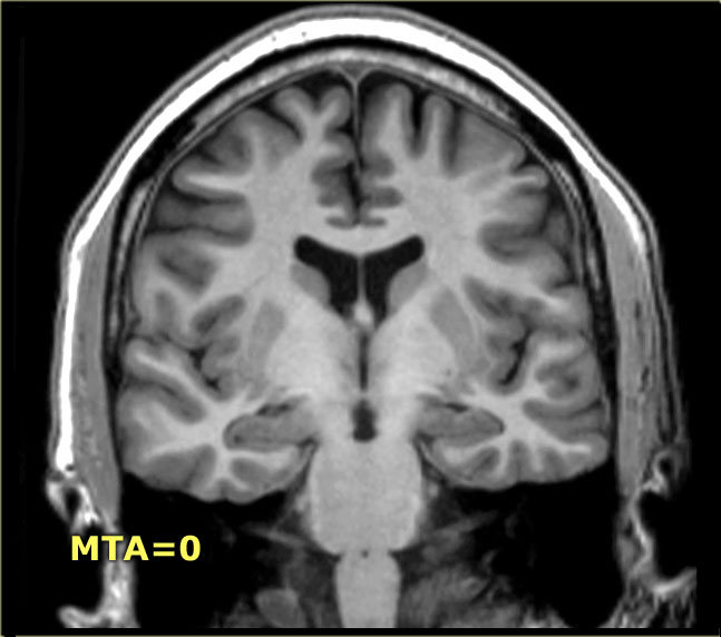
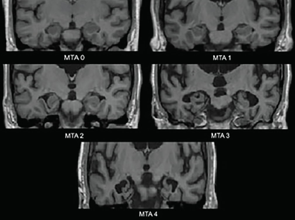
MTA score: 0: no CSF is visible around the hippocampus (normal); 1: choroid fissure is slightly widened; 2: moderate widening of the choroid fissure, mild enlargement of the temporal horn and mild loss of hippocampal height; 3: marked widening of the choroid fissure, moderate enlargement of the temporal horn, and moderate loss of hippocampal hei.

Diagnosis Nerd Radiography Assistants & Imaging Support Workers
Objective To evaluate the diagnostic performance and reliability of the medial temporal lobe atrophy (MTA) scale in patients with Alzheimer's disease. Methods A systematic literature search of MEDLINE and EMBASE databases was performed to select studies that evaluated the diagnostic performance or reliability of MTA scale, published up to January 21, 2021. Pooled estimates of sensitivity and.
The Radiology Assistant Dementia Role of MRI
MTA-scale for Medial Temporal lobe Atrophy Fazekas scale for WM lesions Normal ageing Strategic infarctions Koedam score for Parietal Atrophy FDG-PET Specific Diseases Alzheimers Disease Presenile AD Mild Cognitive Impairment (MCI) Vascular Dementia (VaD) Strategic infarcts and small vessel disease Cerebral Amyloid Angiopathy (CAA)
Demand for Radiologist Assistants Grows as Role Expands
Scores of MTA correlated to age and Mini-mental state examination score. When used to detect DAT from NC, the MTA showed highest diagnostic value than other scales, and when the cutoff score of 1.5 of MTA scale, it obtained an optimal sensitivity (84.5%) and specificity (79.1%) respectively, with a 15.5% of false negative rate.

Image
Abstract. Background: Medial temporal lobe atrophy (MTA) is a sensitive radiologic marker for Alzheimer disease (AD) and associated with cognitive impairment. The value of MTA in the oldest old (>85 years old) is largely unknown.
Careers in Radiologic Technology ASRT
The MTA score, published by Scheltens and colleagues in 1992 [ 4 ], is a simple measure by which mesiotemporal atrophy can be quantified. Using the width of the choroidal fissure, temporal horn, and height of the hippocampal formation, atrophy is evaluated in five grades (0-4).

The Radiology Assistant Brain Dementia Role of MRI
Usage An ERICA score of 2 or 3 (see below) has been shown to have higher diagnostic accuracy for distinguishing healthy controls with subjective cognitive decline from individuals with Alzheimer disease than the older medial temporal lobe atrophy (MTA) score 1 . diagnostic accuracy = 91% sensitivity = 83% specificity = 98% Imaging
Radiologists find their voice in a patientcentric era
Medial temporal lobe atrophy (MTA) is considered as a biomarker for Alzheimer's disease (AD) [ 1 - 6] and visual MTA ratings are available for clinical use [ 7 ]. There is debate as to what cut-off scores should be used in clinical practice to optimally differentiate AD from controls without dementia [ 8] or with other types of dementia [ 9, 10 ].

Medial temporal atrophy (MTA) scoring illustrated on T1weighted MRI.... Download Scientific
Results. Patients with AD had higher MTA scores (mean, 2.13) and ERICA scores (mean, 2.05) than patients with SCD (P < .001).An ERICA score of 2 or greater achieved a higher diagnostic accuracy (91%) than the MTA score (74%), with a sensitivity of 83% versus 57% and a specificity of 98% versus 92% in discriminating dementia caused by AD from SCD (P < .001).

How To A Radiology Assistant Radio Choices
Welcome to the Radiology Assistant. Educational site of the Radiological Society. of the Netherlands. by Frank and Robin Smithuis. Radiology Assistant is a sponsor of Medical Action Myanmar. Horner syndrome. new. Cervical Cancer - MR staging. new.

Radiographs of case 1. (a) The first visit during working length... Download Scientific Diagram
The MTA-score (Scheltens) should be rated on coronal T1-weighted images. on a slice through the corpus of the hippocampus (level of the anterior pons). The scale is based on a visual score of the width of the choroid fissure, the width of the temporal horn, and the height of the hippocampal formation. < 75 years: score 2 or more is abnormal.
Radiology Assistants and Radiology Physician Assistants Collaborative Imaging
Visual rating of medial temporal lobe atrophy (MTA) is often performed in conjunction with dementia workup. Most prior studies involved patients with known or probable Alzheimer's disease (AD). This study investigated the validity and reliability of MTA in a memory clinic population. MTA was rated in 752 MRI examinations, of which 105 were performed in cognitively healthy participants (CH.
Radiologist Assistant (RA) Registered Radiologist Assistant The Radiologic Technologist
Objective To provide age-specific medial temporal lobe atrophy (MTA) cut-off scores for routine clinical practice as marker for Alzheimer's disease (AD). Methods Patients with AD (n = 832, mean age 81.8 years) were compared with patients with subjective cognitive impairment (n = 333, mean age 71.8 years) in a large single-centre memory clinic. Mean of right and left MTA scores was determined.
Imagistica cerebrală în diagnosticul diferențial al demenței Savage Rose
Brain Ischemia. Imaging in Acute Stroke. Vascular territories of the Brain.
