
Pin on Radiopaedia
A subdural hematoma forms because of an accumulation of blood under the dura mater, one of the protective layers to the brain tissue under the calvarium. The understanding of subdural hematoma relies on the knowledge of neuroanatomical sheets covering the brain. The brain is the central repository of delicate neural tissue.
Acute on chronic subdural haematoma Radiology at St. Vincent's University Hospital
Purpose. Subdural hemorrhage (SDH), the accumulation of blood between the dura and arachnoid mater, is one of the most commonly encountered traumatic findings in emergency radiology setting. The purpose of this essay is to review the pitfalls in the diagnosis of SDH including a) mimics on CT imaging and b) etiology other than accidental trauma.
Acute subdural haematoma Radiology at St. Vincent's University Hospital
There are four types of spinal hematomas: epidural, subdural, subarachnoid, and intramedullary (spinal cord) hematomas. Because they differ by their location in relationship to the meningeal membranes and spinal cord, unique radiologic appearances can be recognized to distinguish these types of spinal hemorrhage.
Subdural Hemorrhage in Neonate
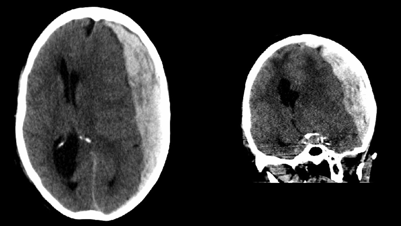
Subdural hemorrhage/hematoma (SDH) is a collection of blood accumulating in the subdural space. Subdural hemorrhage can happen in any age group, is mainly due to head trauma and CT scans are usually sufficient to make the diagnosis. Prognosis varies widely depending on the size and chronicity of the hemorrhage. Epidemiology

Radiology MRI Chronic Subdural Hematoma
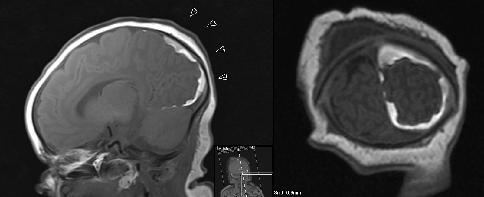
Introduction. Chronic subdural hematoma (cSDH) is a common intracranial hemorrhage, which affects mainly the elderly and is usually caused by trauma ().It is one of the most common conditions in the neurological disciplines (). cSDH is usually diagnosed via non-contrast computed tomography (CT), which is the most common imaging modality due to its sensitivity, widespread availability, and.

Epidural Vs Subdural Hematoma head injury Archives MD direct Treatment of acute subdural
Duret hemorrhages are associated with descending transtentorial herniation, which can occur due to various underlying causes. Herniation syndromes manifest as a result of increased intracranial pressure, leading to shifts in intracranial compartments. The etiology of Duret hemorrhages include 8: epidural hemorrhage. subdural hemorrhage.

Subdural haemorrhage acute Image
Subdural hematoma is a bleeding between the inner layer of the dura mater and the arachnoid mater of the meninges. It usually results from traumatic tearing of the bridging veins that cross the subdural space in patients with anticoagulantia therapy. Epidural hematoma is bleeding in the virtual space between the dura mater and the skull.

Pin on Neuroradiology
Treatment and prognosis EDH is treated with expedient evacuation via a craniotomy. SDH has various management strategies depending on the size, location and extent of mass effect and is either conservative (monitor with serial CT) or surgical (drainage with burr holes). See also EDH EDH (basic article) SDH SDH (basic article) Quiz questions

Rosh Review Subdural hematoma, Emergency medicine, Medical mnemonics
Chronic subdural hematoma (cSDH) is a common intracranial hemorrhage, which affects mainly the elderly and is usually caused by trauma ( 1 ).
Chronic subdural haematoma Radiology at St. Vincent's University Hospital
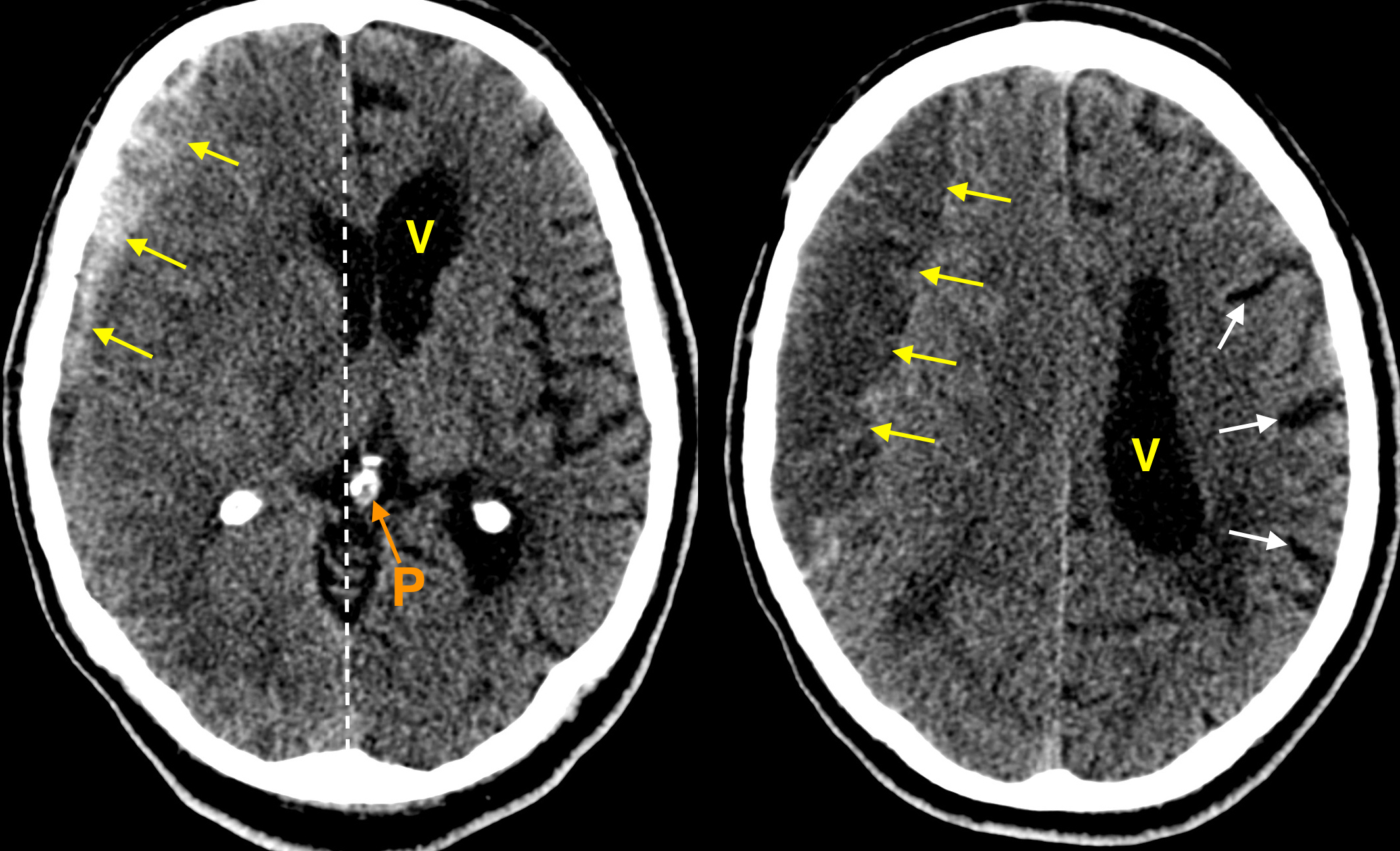
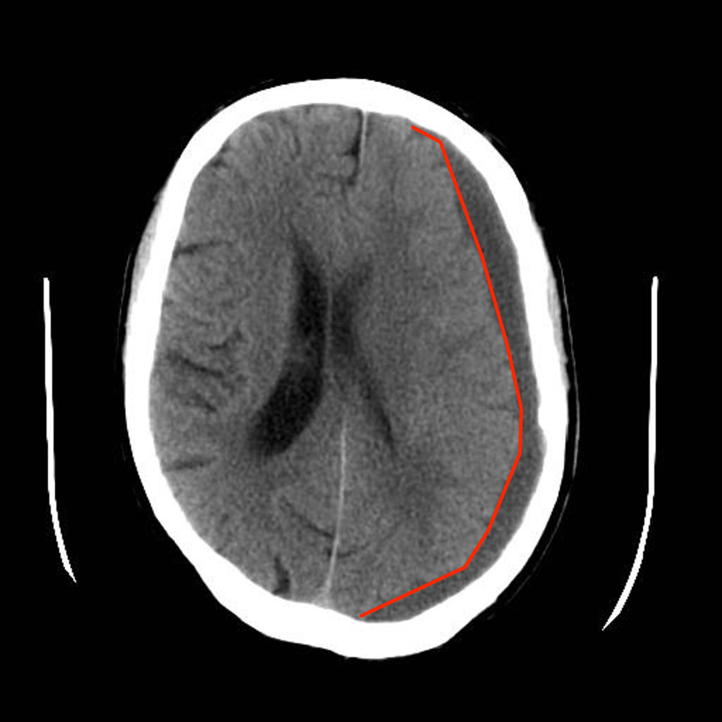
There is a hyperdense right sided extraxial collection measuring up to 20 mm in maximal depth overlying the right cerebral convexity. There is mass effect with approximately 15 mm of midline shift towards the left measured at the level of the third ventricle, with early uncal herniation and obstructive hydrocephalus of the left lateral.
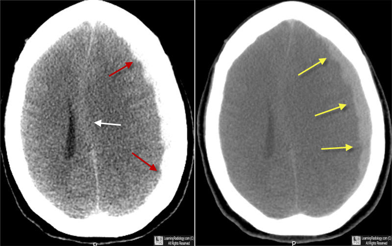
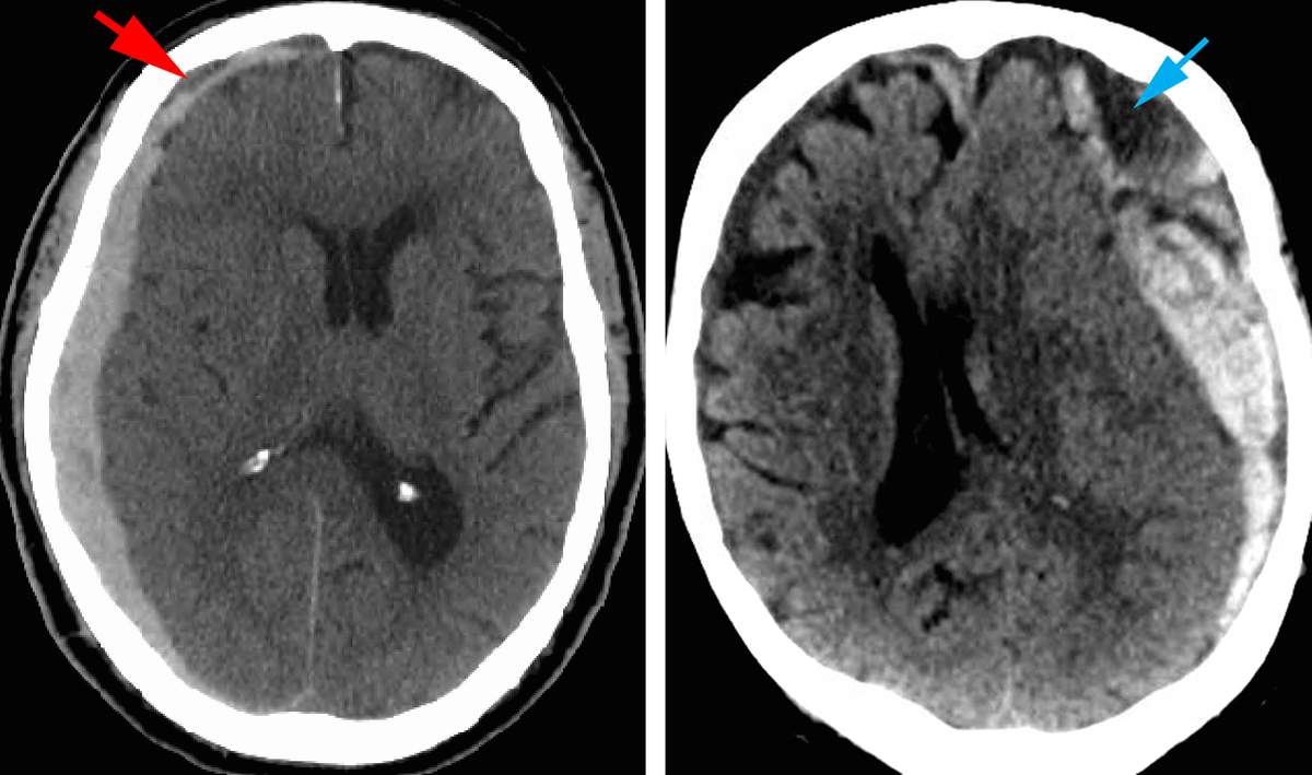
Learning Radiology subdural, hematoma
Intraocular lens (IOL) dislocation is a very rare condition that affects patients who have undergone cataract surgery and consists of the displacement of the implanted lens towards the vitreous cavity of the eye. On other occasions, the lens becomes decentred from the visual axis but does not fall into the vitreous cavity (subluxation).

Subdural Hematoma Neurology Medbullets Step 2/3
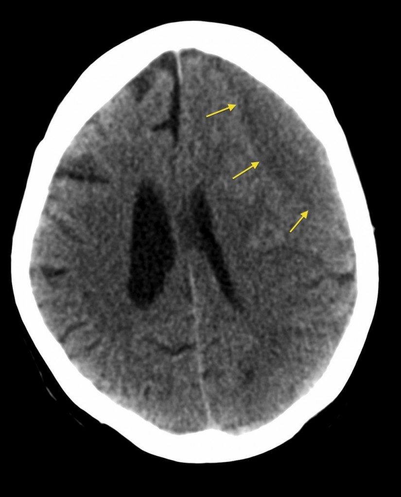
Subdural hemorrhage (SDH) is a collection of blood between the dura and the arachnoid layers of the meninges. They are common and can occur in any age range, usually related to a history of head trauma. Prognosis tends to depend on the extent of the bleed and associated mass effect.
Subdural haematoma Subdural Haemorrhage Geeky Medics
Subdural hematoma (SDH) is a type of bleeding in which a collection of blood gathers between the inner layer of the dura mater and the arachnoid mater of the meninges surrounding the brain. [] Acute SDH is a devastating neurologic injury with significant morbidity and mortality. In patients with large SDH resulting in compression of underlying brain and lateral brain shift, severe neurologic.

Pediatric Subdural Hematoma Pediatric Radiology Reference Article Pediatric Imaging
The oxygenation state of hemoglobin and its location (whether it is contained within red blood cells or diffused in the extracellular space) have a tremendous effect on the imaging effects of blood. The three hemoglobin states to be considered are oxyhemoglobin, deoxyhemoglobin and methemoglobin.
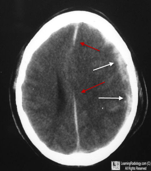
Learning Radiology subdural, hematoma
A subdural hematoma is a type of bleed inside your head. It's a type of bleed that occurs within your skull but outside the actual brain tissue. The brain has three membrane layers or coverings (called meninges) that lie between the bony skull and your brain tissue. The purpose of the meninges is to cover and protect the brain.
Acute Subdural Hematoma The Neurosurgical Atlas
Chronic subdural hematoma (CSDH), which generally occurs in elderly patients, is a frequently diagnosed condition in neurosurgical departments. Computed tomography (CT) and magnetic resonance imaging (MRI) are the most preferred diagnostic modalities for CSDH assessment.
