
Internal Female Anatomy Superior View TrialExhibits Inc.
Female Anatomy Diagrams of the inside and outside of female body parts By Brandi Jones, MSN-ED RN-BC Updated on April 26, 2023 Medically reviewed by Lauren Schlanger, MD Fact checked by Sarah Scott Table of Contents View All Diagram External Internal Breast Anatomy Functions
Abdomen AnatomyFemale Female Abdominal Anatomy Illustration Stock Image F026 5545 Science
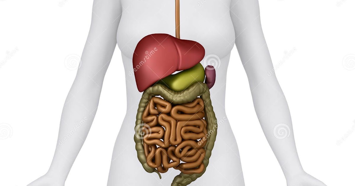
The abdomen is the part of the body that contains all of the structures between the thorax (chest) and the pelvis, and is separated from the thorax via the diaphragm. The region occupied by the abdomen is called the abdominal cavity, and is enclosed by the abdominal muscles at front and to the sides, and by part of the vertebral column at the back.

Abdomen Anatomy Female / Anatomy Of Female Abdomen And Pelvis Trialexhibits Inc
Reading time: 17 minutes Recommended video: Surface anatomy of the abdomen and the lower extremity [13:14] Overview of the surface anatomy landmarks found in the abdomen and lower limbs. Abdomen 1/2 Synonyms: Abdominal region, Regio abdominis , show more. Hello there fellow anatomist and welcome to abdomen and pelvis 101!
Anatomy Of The Female Abdomen And Pelvis, Cut away View Healthiack
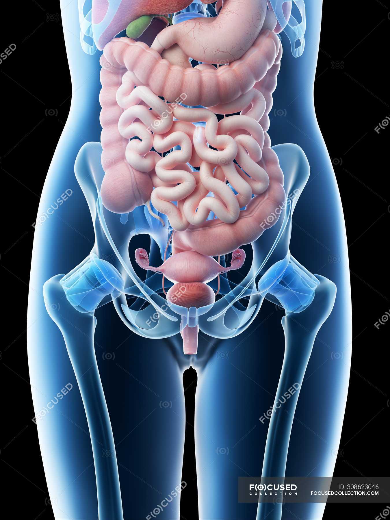
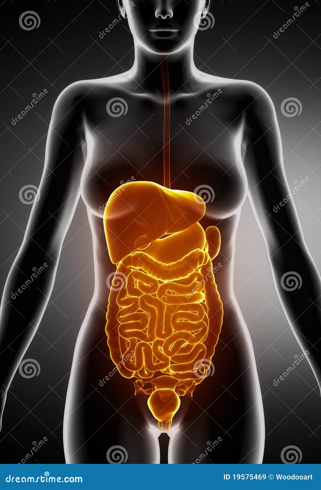
This medical exhibit diagram illustrates the anatomy of the female abdomen and pelvis from an anterior (front) cut-away view, showing elements of the digestive system. The liver, stomach, and abdominal contents are clearly identified and labeled, including the cecum, ascending colon, transverse colon, descending colon, and small intestine.
Abdomen Anatomy Female Body Illustration of female digestive system back view Stock Photo
The abdomen (colloquially called the belly, tummy, midriff, tucky or stomach) is the part of the body between the thorax (chest) and pelvis, in humans and in other vertebrates.The abdomen is the front part of the abdominal segment of the torso.The area occupied by the abdomen is called the abdominal cavity.In arthropods, it is the posterior tagma of the body; it follows the thorax or.
Female Abdominal Anatomy Pictures / Stock Images Female Abdominal Organs Stock Photography
Anatomy atlas of the female pelvis: 101 labeled illustrations of the female genital system (ovaries, uterine tubes, uterus, vagina, vulva, clitoris) and pelvic cavity (bladder, rectum, pelvic diaphragm, perineum with innervation and blood supply). Tome 2 : Thorax, coeur, abdomen et pelvis. Torsten B. Möller - Emil Reif. Paru le : 06/2014.

Abdominal Anatomy Pictures Female Female Human Body Organs Diagram Don't
Structure and Function The uterus sits in the center of the female pelvic cavity (Figure 2.) The most common position of the uterus in the pelvic cavity is anteverted and anteflexed. [1] " Version" refers to the angle between the cervix and the vagina. An anteverted uterus appears "tipped forward" in the pelvic cavity.

Abdomen Wikipedia, la enciclopedia libre
Below is a 3D model of female anatomy, which is fully interactive. Explore the model using your mouse pad or touchscreen to understand more about female anatomy. External anatomy The external.

Female Abdominal Anatomy TrialExhibits Inc.
This medical illustration depicts a mid-sagittal view of the normal anatomy of the female abdomen and pelvis. Labeled structures include the large bowel (colon or large intestine), umbilicus, small intestine, ovary, fallopian tube, uterus and bladder. Variations Anatomia do Abdômen e da Pélvis Feminina - exh6130apt-br
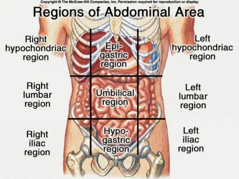
Female Abdomen Anatomy Quadrants / Abdominal Surface Anatomy Radiology Reference Article
The abdominal muscles stretch during pregnancy to allow room for the fetus to grow. The rapid growth can sometimes cause stretch marks on the skin, but these can be prevented or reversed with.

Female Anatomy Upper Body Stock Photo Download Image Now iStock
Diagram of Female Abdomen: Understanding the Anatomy and Functions. The female abdomen is a complex and vital part of the body. Understanding its anatomy and functions can help women take better care of their abdominal health. In this blog post, we will explore the diagram of the female abdomen, highlighting its various structures and their.

Female abdominal anatomy, illustration Stock Image F026/5545 Science Photo Library
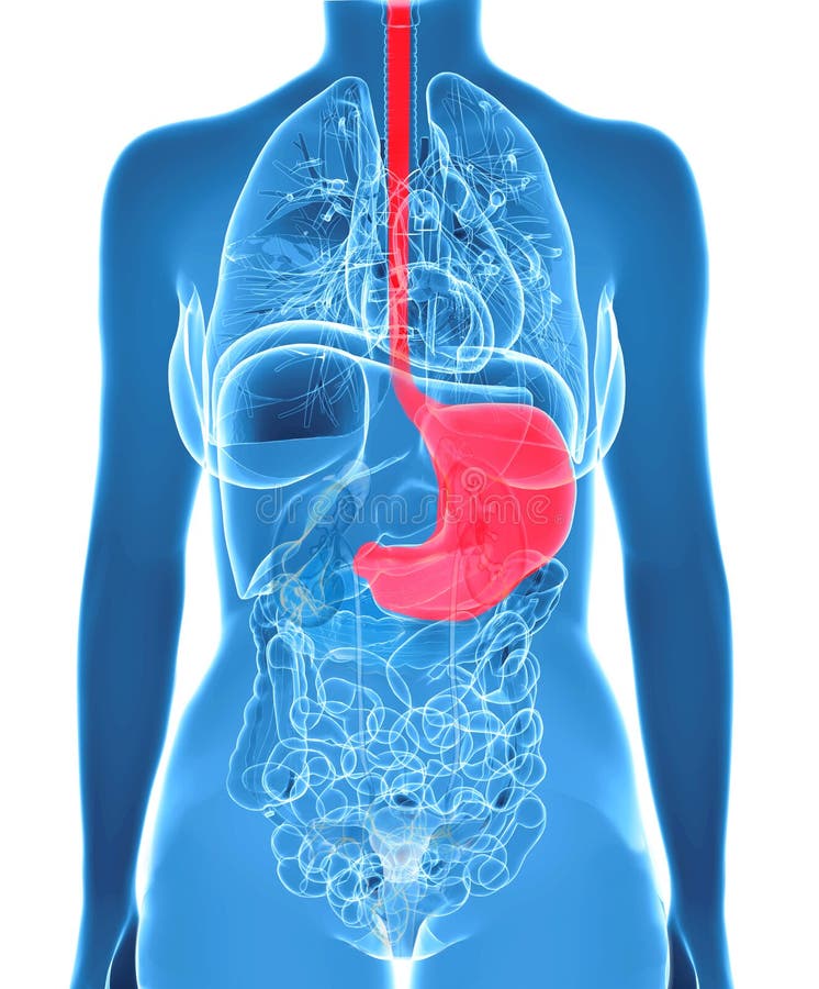
1. Anterior view: anatomy of female abdomen and pelvis: skin. 2. Anterior view: anatomy of female abdomen and pelvis: muscles of anterior abdomen wall. 3. Anterior view: anatomy of female abdomen and pelvis: stomach and omentum. 4. Anterior view: anatomy of female abdomen and pelvis: small bowel and colon. 5.
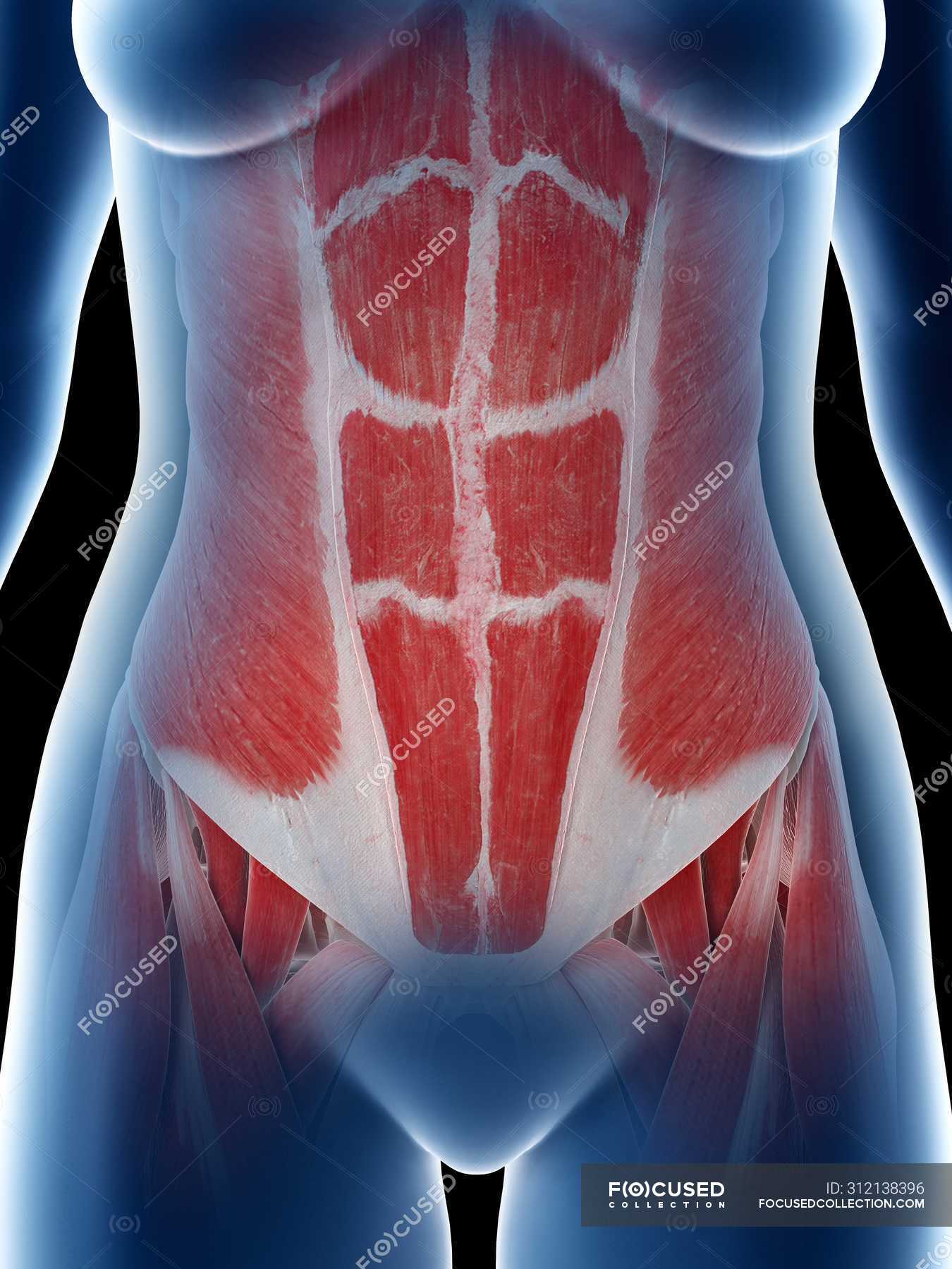
Female Abdomen Muscle Anatomy Abdomen And Pelvis Female Stock Photo Alamy The muscles fibers
Picture of Abdomen The abdominal cavity is the part of the body that houses the stomach, liver, pancreas, kidneys, gallbladder, spleen, and the large and small intestines. The diaphragm marks the top of the abdomen and the horizontal line at the level of the top of the pelvis marks the bottom.
Female Abdominal Organs Posterior View Stock Image 19746049
ISSN 2534-5079. This e-Anatomy illustrates the gross anatomy of the digestive system. We focused especially on the diagrams of the abdominal digestive system (oesophagus is described on the modules about the thorax and oral cavity/pharynx on the ENT modules). 84 anatomical diagrams and histological images with over 300 labeled anatomical parts.
:max_bytes(150000):strip_icc()/abdominal-muscle--illustration-687796219-5bfd930046e0fb00269d5c12.jpg)
Muscles In Lower Left Abdomen Left Vs Right Back And Abdominal Pain In Women oceaniclore
Female Pelvis Overview Anatomy and function Diagram Conditions Symptoms Health tips What is the female pelvis? The pelvis is the lower part of the torso. It's located between the abdomen.

Human Anatomy Abdomen Stomach Pics Anatomy organs, Animal cell anatomy, Human anatomy
Muscular System Organs and Inner Muscles Organs and Inner Muscles The pelvic region holds major organs under its layers of muscles. Some of the most important include the major digestive.
