How to Use a Microscope
Microscope Parts and Functions With Labeled Diagram and Functions How does a Compound Microscope Work?. Before exploring microscope parts and functions, you should probably understand that the compound light microscope is more complicated than just a microscope with more than one lens.. First, the purpose of a microscope is to magnify a small object or to magnify the fine details of a larger.

PPT Label the parts on your microscope picture. PowerPoint Presentation ID1888029
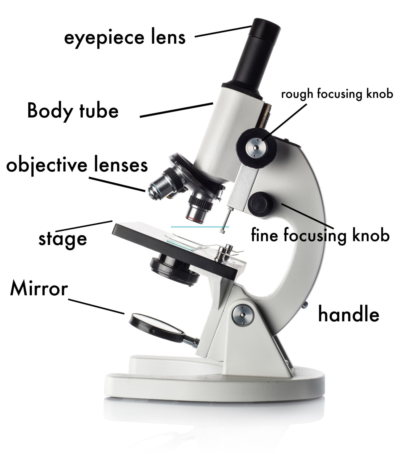
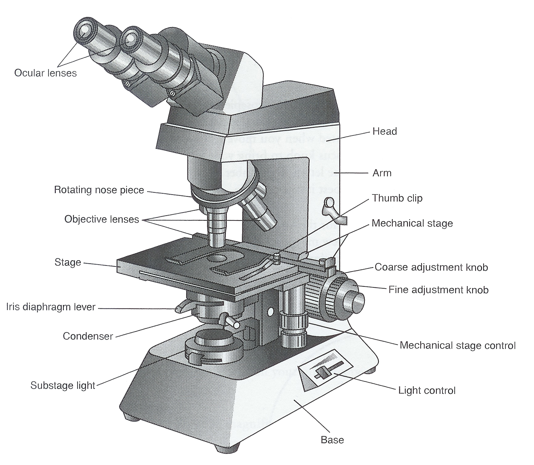
Tube: Connects the eyepiece to the objective lenses. Arm: Supports the tube and connects it to the base. Base: The bottom of the microscope, used for support. Illuminator: A steady light source (110 volts) used in place of a mirror. If your microscope has a mirror, it is used to reflect light from an external light source up through the bottom.

Microscope Labeling
Microscopes are instruments that are used in science laboratories to visualize very minute objects, such as cells and microorganisms, giving a contrasting image that is magnified. Microscopes are made up of lenses for magnification, each with its own magnification powers.

Microscope Diagram to Print 101 Diagrams
Label the microscope Interactive Add to collection Use this interactive to identify and label the main parts of a microscope. Drag and drop the text labels onto the microscope diagram. eye piece lens coarse focus adjustment base stage diaphragm or iris high-power objective light source fine focus adjustment Download Exercise Tweet
Microscope Drawing Template at GetDrawings Free download
" Micro " means very small (typically not visible to the naked eye) and " scope " means to assess or investigate carefully. So, the microscope is an instrument that aids users to carefully investigate and assess microscopic organisms and objects that are not visible to the naked eye. Table of Contents Simple Microscope Compound Microscope

Label And Color The Parts Of Both Microscopes Ythoreccio
Microscope Description. A microscope is a laboratory instrument used to examine objects that are too small to be seen by the naked eye. In other words, it enlarges images of small objects. Invented by a Dutch spectacle maker in the late 16th century, light microscopes use lenses and light to magnify images.

Microscope Diagram Labeled, Unlabeled and Blank Parts of a Microscope
Magnification is a measure of how much larger a microscope (or set of lenses within a microscope) causes an object to appear. For instance, the light microscopes typically used in high schools and colleges magnify up to about 400 times actual size. So, something that was 1 mm wide in real life would be 400 mm wide in the microscope image.
Monday September 25 Parts of a Compound Light Microscope
A microscope is a piece of laboratory optical equipment used to magnify small things that are too small for the details to be seen by the naked eye. The microscope is the microbiologist's most basic tool, and every microbiology student needs some background knowledge on parts of a microscope and how microscopes work.

Parts of the Microscope with Labeling (also Free Printouts)
Download the Label the Parts of the Microscope PDF printable version here. Download the Label the Parts of the Microscope: Answers PDF printable version here. Microscope World explains the parts of the microscope, including a printable worksheet for schools and home.

LABEL MICROSCOPE PARTS « Optical Instruments
A light microscope is a biology laboratory instrument or tool, that uses visible light to detect and magnify very small objects and enlarge them. They use lenses to focus light on the specimen, magnifying it thus producing an image. The specimen is normally placed close to the microscopic lens.
Light Microscope Main Parts Of Light Microscope Biology —
The most familiar type of microscope is the optical, or light, microscope, in which glass lenses are used to form the image. Optical microscopes can be simple, consisting of a single lens, or compound, consisting of several optical components in line. The hand magnifying glass can magnify about 3 to 20×. Single-lensed simple microscopes can.

Microscope Interactive worksheet
Open combination drawer and take out the microscope. 3. Label all the parts of the microscope with the provided post-its using the image below or the laboratory manual. Note. The image below does not match your microscope perfectly, you will be responsible for knowing the parts of your microscope on the lab practical.
35 Label Of Compound Microscope Labels 2021
There are 1000 millimeters (mm) in one meter. 1 mm = 10 -3 meter. There are 1000 micrometers (microns, or µm) in one millimeter. 1 µm = 10 -6 meter. There are 1000 nanometers in one micrometer. 1 nm = 10 -9 meter. Figure 1: Resolving Power of Microscopes. The microscope is one of the microbiologist's greatest tools.
Ag Biology Unit 2
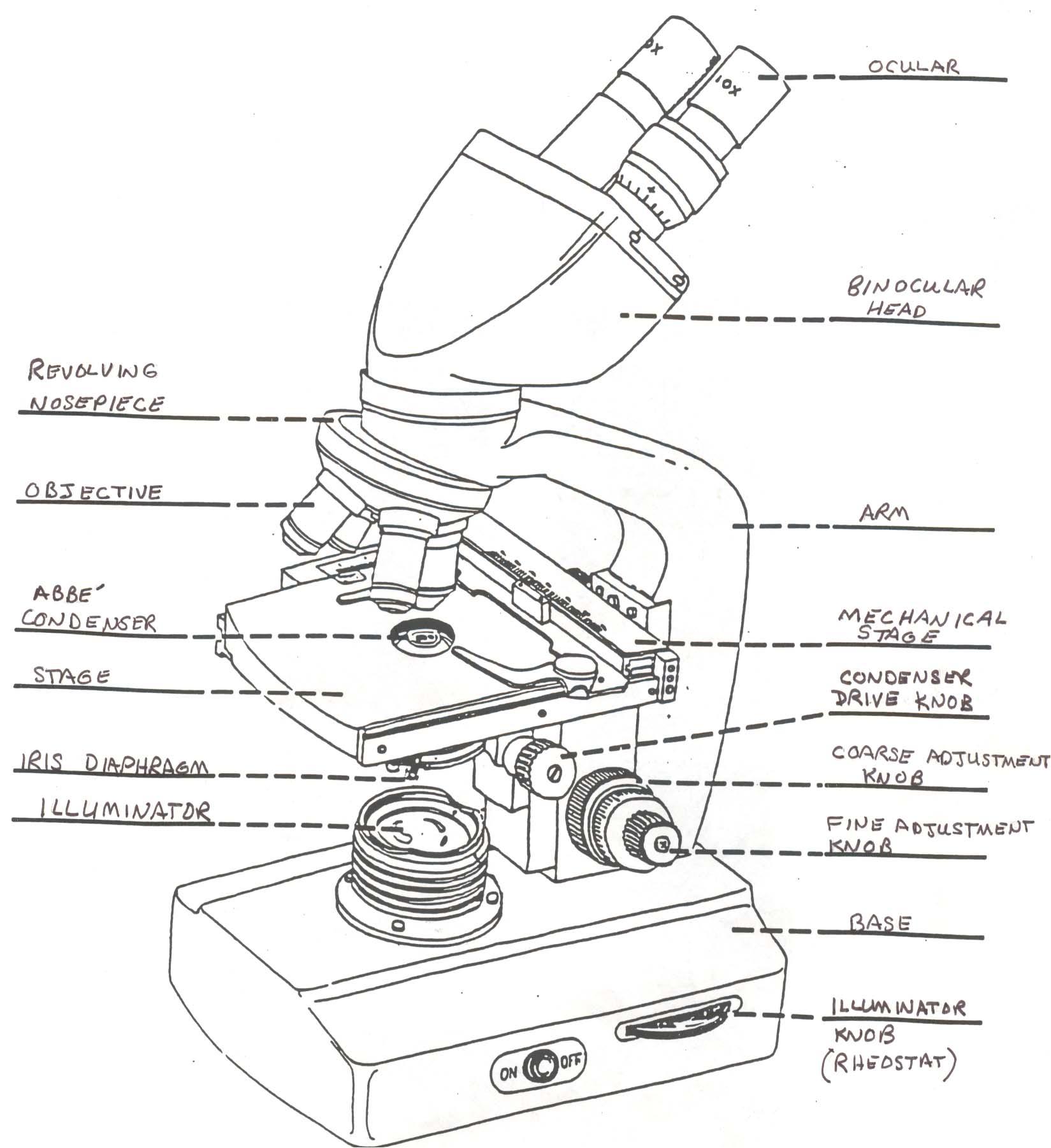
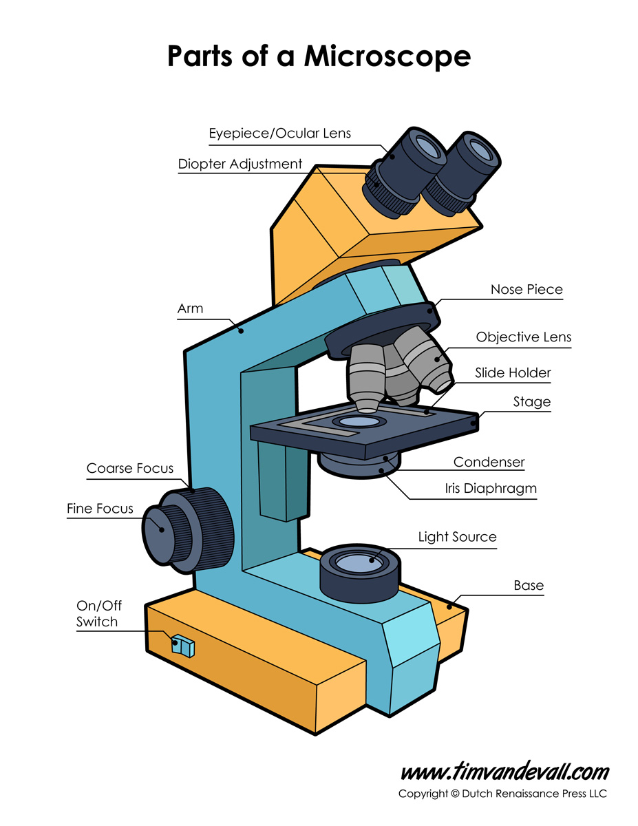
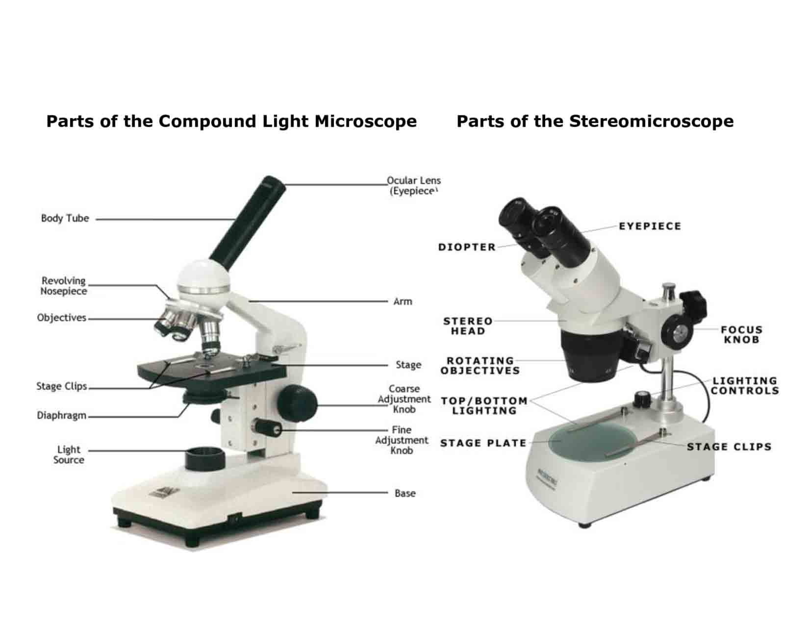
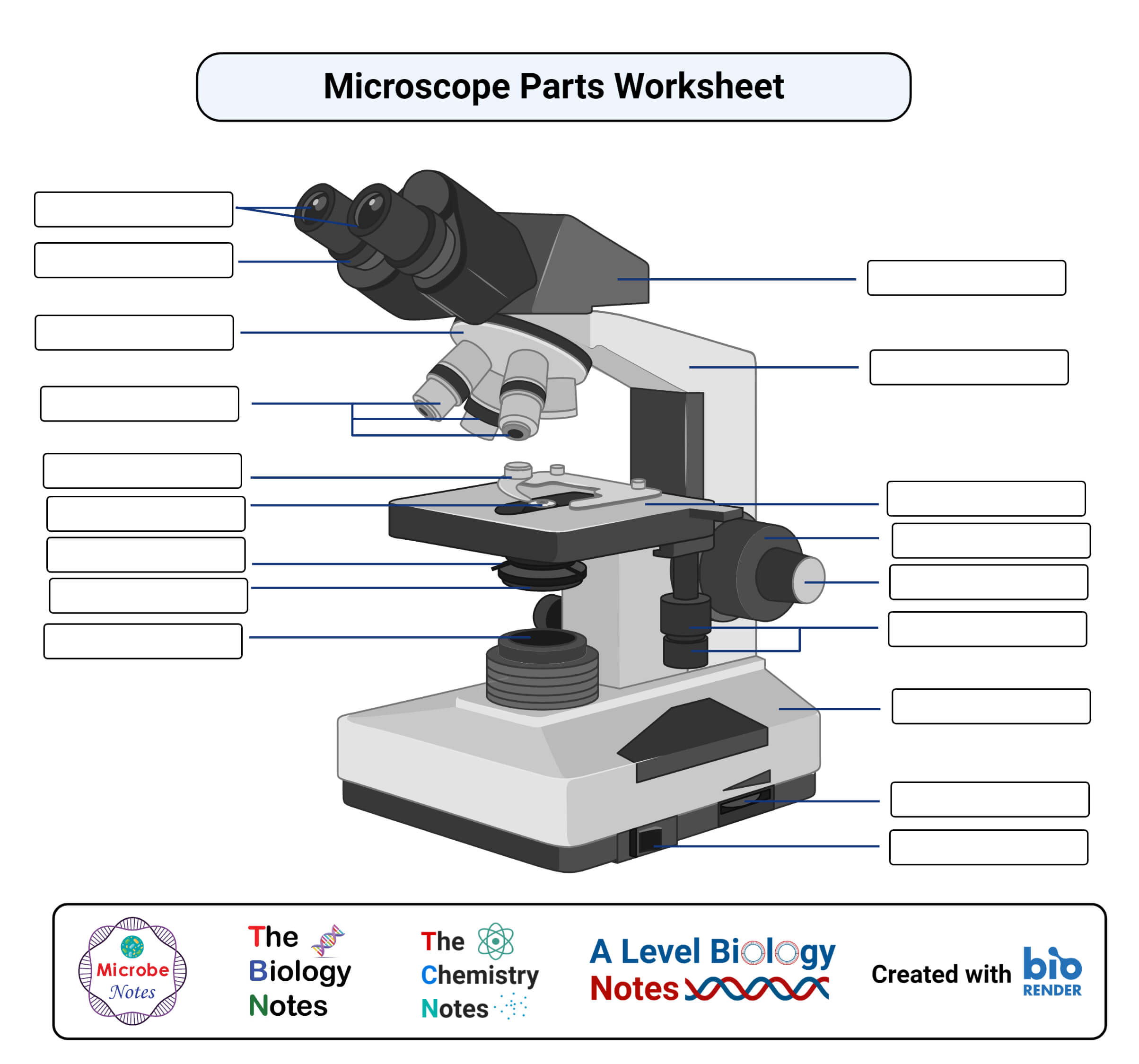
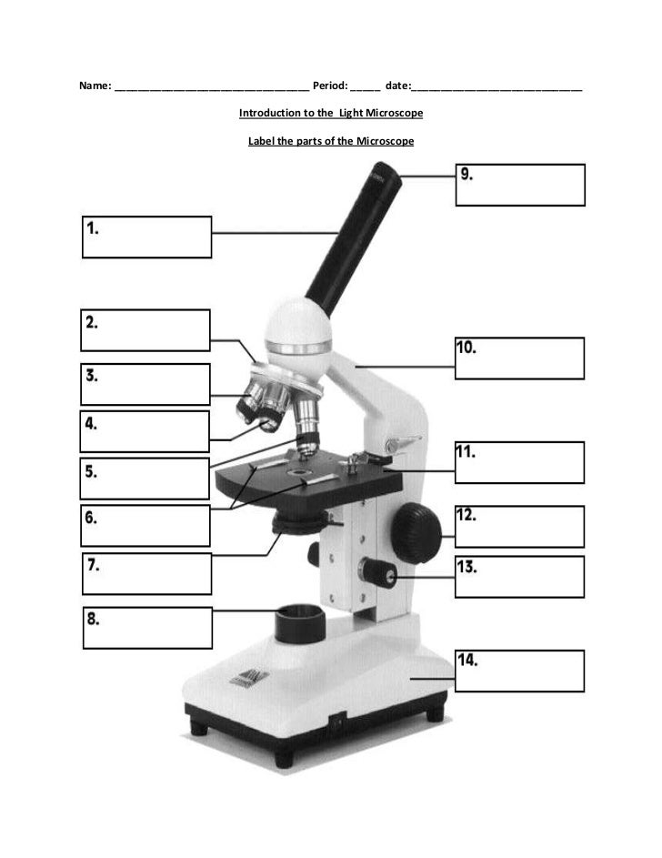
1. Eyepiece 2. Body tube/Head 3. Turret/Nose piece 4. Objective lenses 5. Knobs (fine and coarse) 6. Stage and stage clips 7. Aperture 9. Condenser 10. Condenser focus knob 11. Iris diaphragm 12. Diopter adjustment 13. Arm 14. Specimen/slide 15. Stage control/stage height adjustment 16. On and off switch 17. Base

This is a common compound microscope. Label its pa
The labeling worksheet could be used as a quiz or as part of direct instruction. Students label the microscope as you go over what each part is used for. Then they answer short fill in the blank sentences about the proper use of the microscope. The google slides shown below have the same microscope image with the labels for students to copy.
Biology label part of microscope
"Micro" means small or tiny. "Scope" means to view or to observe. Therefore, a microscope can be understood as an instrument to see tiny things. What is a "compound microscope"? A compound microscope is the most common type of light (optical) microscopes. The term "compound" refers to the microscope having more than one lens.
