19 Best Cow Eye Dissection Labeled
Cow's Eye Dissection - How does your eye work? You see the world because light gets into your eyes. Your eye uses that light to make an image of the world inside your eye—just as a camera uses light to make a photograph. To understand how your eye makes an image of the world, you need to know a little bit about lenses.

Cow Eye Diagram Quizlet
Cow eye diagrams can be used to teach farmers and veterinarians about the signs and symptoms of these diseases, as well as the best treatment options. In addition to disease and injury, the cow eye is also affected by environmental factors such as light and temperature. Cow eye diagrams can be used to teach students about the different.
19 Best Cow Eye Dissection Labeled
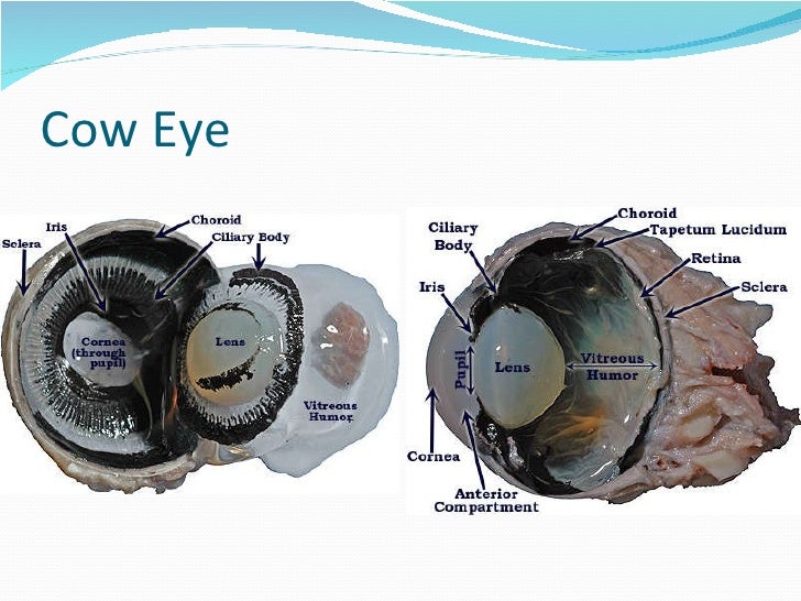
Updated April 24, 2018 By Alison Cooksey The eyeballs of humans and the eyeballs of cows have a similar structure overall. Both have the sclera, which is the white part of the eyeball, cornea or the clear structure over the iris and pupil, lens, vitreous fluid, retina and choroid.
Cow Eye Labeled Diagram ClipArt Best
The cow eye is a fantastic specimen for students of all ages to dissect. The structures are clear, dissection easy to accomplish and usually kids enjoy the lab.. Plus, I've found that my anatomy students have trouble matching parts on models to the real thing. I also leave diagrams on lab tables to help locate structures. Common Hurdles.

Cow Eye Model Diagram Quizlet
Download step-by-step instructions (PDF file) for doing your own cow's eye dissection. Instructions include an eye diagram, a glossary, and color photos for each step.
Cow Eye Labeled ClipArt Best
Start studying External and internal anatomy of the cow eye. Learn vocabulary, terms, and more with flashcards, games, and other study tools.
Cow Eye Labeled Diagram ClipArt Best
Learn how to dissect a cow's eye in your classroom. This resource includes: a step-by-step, hints and tips, a cow eye primer, and a glossary of terms. Cow's Eye Dissection - Eye diagram

Cow's Eye Dissection Instructions
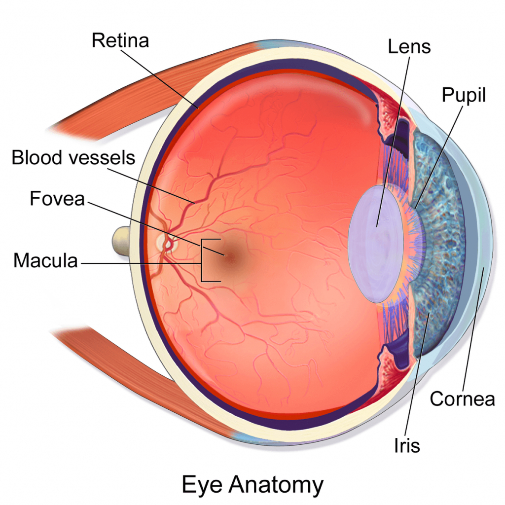
cornea. Clear, outer layer of the front of the eye. sclera. White, outermost layer of the eye. Helps maintain shape and gives attachment to muscles. photoreceptors. The cells in the retina that respond to light (rods and cones) rods. Photoreceptor cells in the eye that detect black, white, and gray.

Cow Eye Dissection Parts Labeled All About Cow Photos
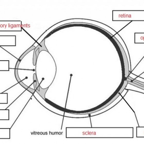
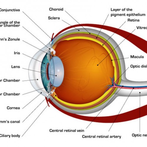
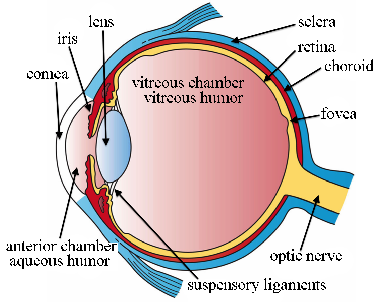
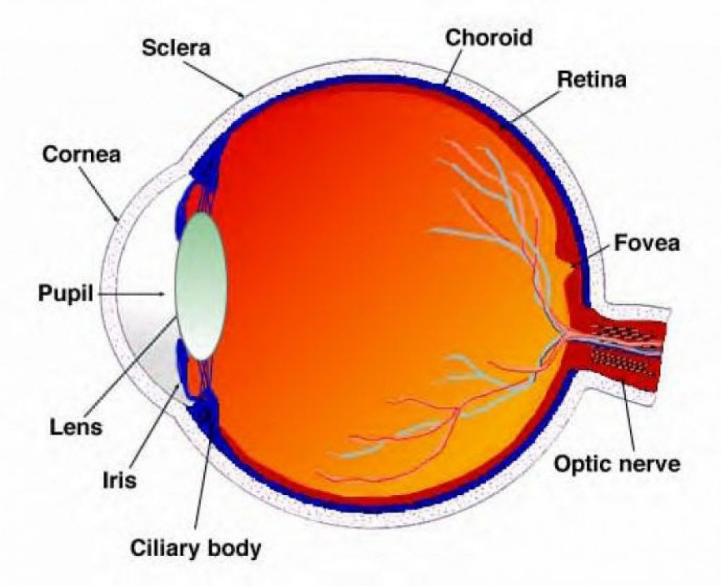
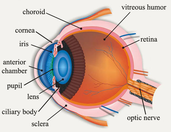
This diagram shows the parts of the eye. Can you find these parts in a cow's eye? Here's what you do: Examine the outside of the eye. See how many parts of the eye you can identify. You should be able to find the whites (or sclera), the tough, outer covering of the eyeball.
Cow Eye Labeled ClipArt Best
Cow Eye Dissection. The mammalian eye is a sensory organ that operates as part of the nervous system. These complex organs gather light, focus it on receptor cells, and transmit the information to the brain where it is interpreted. Placement and shape of eyes vary across the animal kingdom, but the main function remains consistent—vision.

Cow's Eye Dissection Eye diagram Home School Science Pinterest
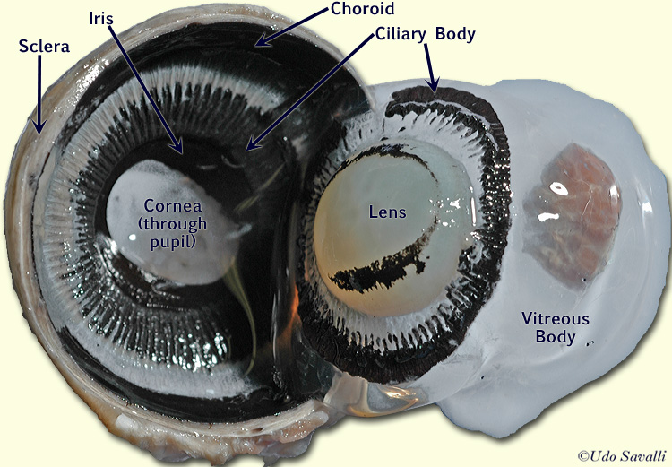
Diagram of the Eye. Procedure. 1. Cut the fat around the eye. 2. Puncture the cornea with scissors or a scalpel. The cornea is pinned here. 3. Cut the eye into a front and a back half. 4. Open the eye. The gelatinous fluid inside is the vitreous humor, the lens sits within this liquid. 5. Separate the parts of the eye.
Cow Eye Labeled Diagram ClipArt Best
In the cow's eye dissection, we cut away all the fat and muscle so that we can see the eyeball. Step 4: The cornea protects the eye.. Printable diagram. Experimenting with a Lens. To understand how your eye makes an image of the world, you need to know a little bit about lenses. Learn about lenses and experiment with a magnifying glass to.

Gross Anatomy Of Cow Eye ANATOMY
Look down. Look all around. Six muscles attached to your eyeball move your eye so you can look in different directions. Cows have only four muscles that control their eyes. They can look up, down, left, and right, but they can't roll their eyes like you can. WATCH VIDEO
Cow Eye Labeled
1. Examine the outside of the eye. You should be able to find the sclera, or the whites of the eye. This tough, outer covering of the eyeball has fat and muscle attached to it 2. Locate the covering over the front of the eye, the cornea. When the cow was alive, the cornea was clear. In your cow's eye, the cornea may be cloudy or blue in color. 2.
human eye colour diagram Clip Art Library
LAB 13 EXERCISE 13.7. 1. 1. Examine the outside of the eye. You should be able to find the sclera, or the whites of the eye. This tough, outer covering of the eyeball has fat and muscle attached to it. 2. Locate the covering over the front of the eye, the cornea. When the cow was alive, the cornea was clear.

Cow Eye Diagram Quizlet
Clear protective covering over the front of eye that bends light entering eye. Pupil. The hole where light passes into the lens. Iris. The colored part of the eye that regulates the size of the pupil to control light entering the eye. Vitreous Humor. A clear liquid inside eyeball that gives the eye its round shape. Lens.
